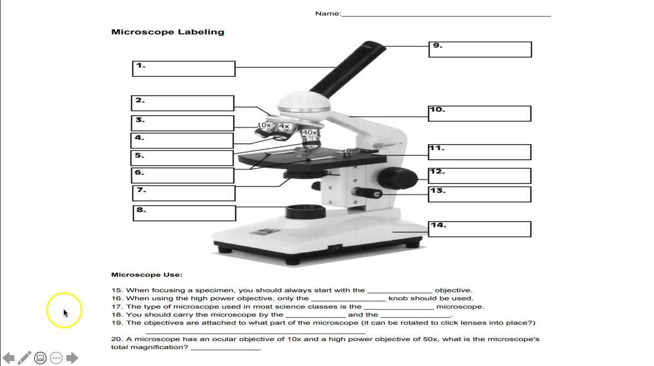
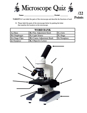
44 microscope diagram without labels
How to run an assay | Agilent Place the plate in a 37°C incubator without CO 2 for one hour prior to the assay. Remove all but 50 μL of the culture medium from each well. The small amount of medium is left to keep the cells from drying out. Gently add 1mL of assay medium. Place the plate in a 37°C incubator without CO 2 for one hour prior to the assay. Microscope Parts, Types & Diagram | What is a Microscope? Microscope Diagram There are many illustrations available for the diagram of a light microscope. The essential parts include the head, base, arms, lenses, and lights. In diagrams, one...
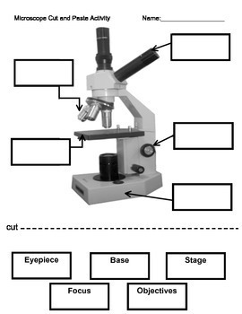
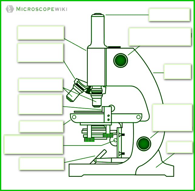
Labeling the Parts of the Microscope | Microscope World Resources Labeling the Parts of the Microscope This activity has been designed for use in homes and schools. Each microscope layout (both blank and the version with answers) are available as PDF downloads. You can view a more in-depth review of each part of the microscope here. Download the Label the Parts of the Microscope PDF printable version here.

Microscope diagram without labels
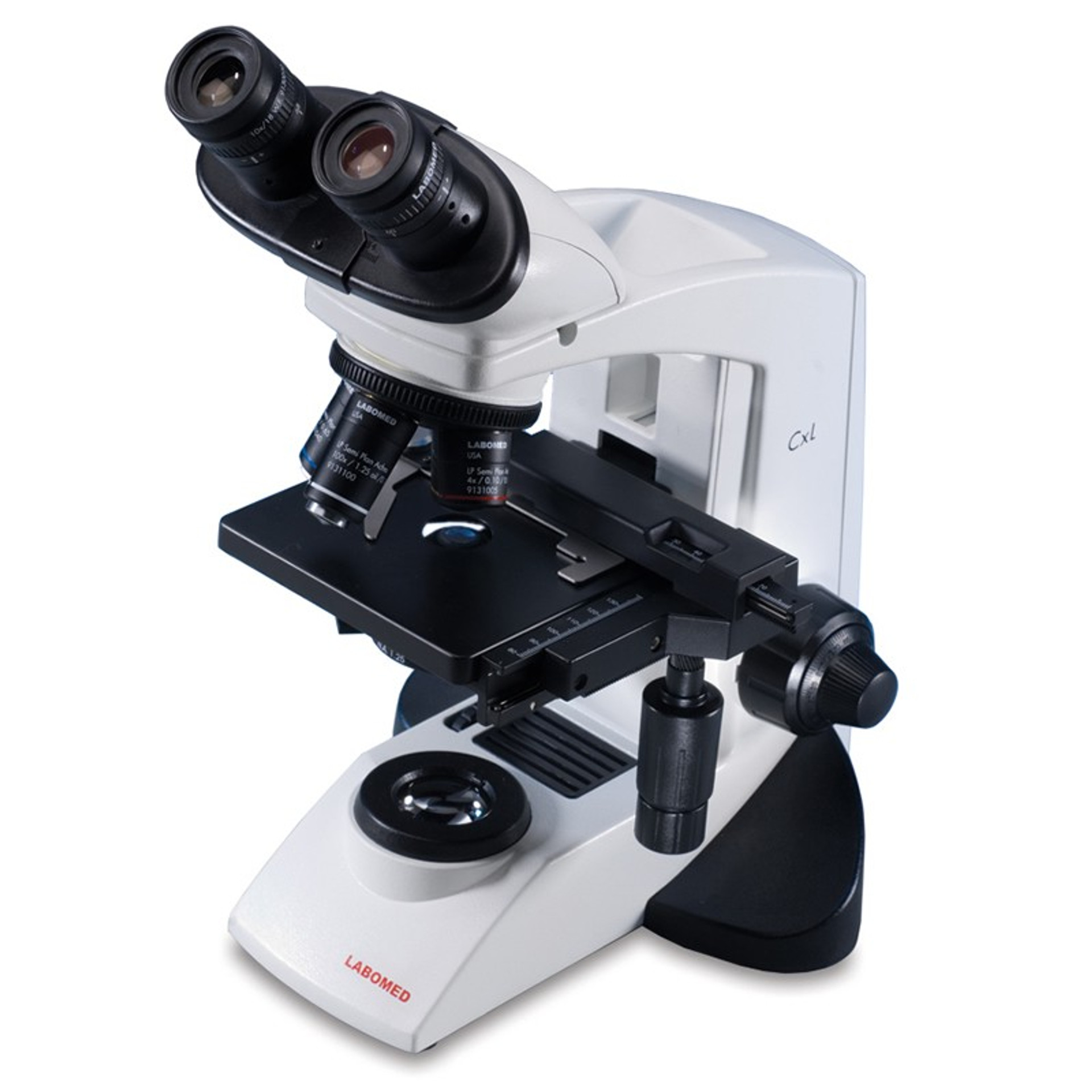
Compound Microscope Parts, Functions, and Labeled Diagram Nov 18, 2020 · Common compound microscope parts include: Compound Microscope Definitions for Labels Eyepiece (ocular lens) with or without Pointer: The part that is looked through at the top of the compound microscope. Eyepieces typically have a magnification between 5x & 30x. How does a Microscope work A compound microscope has two or more lenses. The eyepiece or ocular lens sits atop the body tube. Many microscopes are binocular and have two ocular lenses. Additionally, a binocular head will have a prism, either in the head or the body tube, to split the image and direct it to both oculars. Wikipedia:Citation needed - Wikipedia To ensure that all Wikipedia content is verifiable, Wikipedia provides a means for anyone to question an uncited claim.If your work has been tagged, please provide a reliable source for the statement, and discuss if needed.. You can add a citation by selecting from the drop-down menu at the top of the editing box.In markup, you can add a citation manually using ref tags.
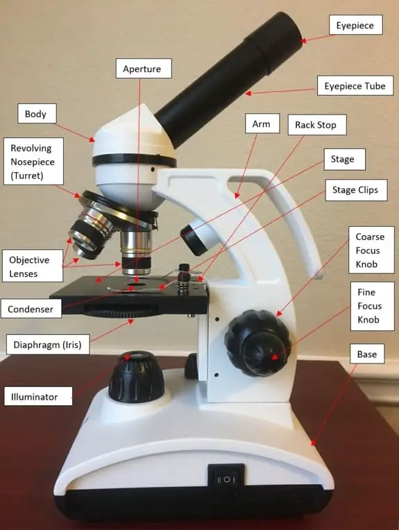
Microscope diagram without labels. Microscope Types (with labeled diagrams) and Functions Phase-contrast microscope labeled diagram Phase-contrast microscope functions: Its applications areas include In cases where the specimen is colorless and is very tiny In biology to conduct cellular level examination of microorganisms that can't be visualized using the bright field microscopy Interference Microscope Lipid bilayer - Wikipedia The lipid bilayer is very thin compared to its lateral dimensions. If a typical mammalian cell (diameter ~10 micrometers) were magnified to the size of a watermelon (~1 ft/30 cm), the lipid bilayer making up the plasma membrane would be about as thick as a piece of office paper. Despite being only a few nanometers thick, the bilayer is composed of several distinct chemical … Microscope, Microscope Parts, Labeled Diagram, and Functions The Iris Diaphragm is located above the condenser lens and below the microscope stage. The different sized holes in the diaphragm helps to vary the size of the cone and intensity of light that is projected upward into the slide. However, there is no set rule regarding which setting to use for a particular power. Parts of a microscope with functions and labeled diagram - Microbe Notes Figure: Diagram of parts of a microscope There are three structural parts of the microscope i.e. head, base, and arm. Head - This is also known as the body. It carries the optical parts in the upper part of the microscope. Base - It acts as microscopes support. It also carries microscopic illuminators.
Join LiveJournal Password requirements: 6 to 30 characters long; ASCII characters only (characters found on a standard US keyboard); must contain at least 4 different symbols; Catalyst support effects on hydrogen spillover | Nature Jan 05, 2017 · The mechanism of hydrogen spillover is described using a precisely nanofabricated model system, explaining why it is slower on an aluminum oxide catalyst support than on a titanium oxide catalyst ... Microscope Diagram - Label Diagram | Quizlet The bottom of the microscope, used for support. ocular lens. Eyepiece of a microscope. Diaphragm. Regulates the amount of light on the specimen. nosepiece of microscope. holds the objective lenses. objective lens. The lens on a light microscope that is closest to the stage. Flexography - Wikipedia Flexography (often abbreviated to flexo) is a form of printing process which utilizes a flexible relief plate. It is essentially a modern version of letterpress, evolved with high speed rotary functionality, which can be used for printing on almost any type of substrate, including plastic, metallic films, cellophane, and paper.It is widely used for printing on the non-porous substrates ...
Binocular Microscope Anatomy - Parts and Functions with a Labeled Diagram Now, I will discuss the details anatomy of the light compound microscope with the labeled diagram. Why it is called binocular: because it has two ocular lenses or an eyepiece on the head that attaches to the objective lens, this ocular lens magnifies the image produced by the objective lens. Binocular microscope parts and functions Label the microscope — Science Learning Hub In this interactive, you can label the different parts of a microscope. Use this with the Microscope parts activity to help students identify and label the main parts of a microscope and then describe their functions. Drag and drop the text labels onto the microscope diagram. List of Top 7 Types of Microscopes (With Diagram) - Biology Discussion ADVERTISEMENTS: List of top seven types of microscopes:- 1. Phase Contrast Microscope 2. Interference Contrast Microscope 3. Ultraviolet Microscope 4. Fluorescence Microscope 5. Immunofluorescence 6. Dark-Field Microscope 7. Electron Microscope. Type # 1. Phase Contrast Microscope: This microscope was developed by Fritz Zernikes (1935), a Dutch physicist who was awarded Nobel Prize in 1953 ... Simple Microscope - Parts, Functions, Diagram and Labelling A simple microscope is a device that only has one lens for magnification. It functions the same way as the magnifying glass. Although it is simple in terms of design and function, it is useful I various fields including medicine, jewelry and watchmaking, and agriculture, to name a few. References
Microscope Diagram - Label Diagram | Quizlet
High-density switchable skyrmion-like polar nanodomains Mar 02, 2022 · Topological domains in ferroelectrics1–5 have received much attention recently owing to their novel functionalities and potential applications6,7 in electronic devices. So far, however, such ...

Parts of a Microscope Labeling Activity
Compound Microscope Parts - Labeled Diagram and their Functions The eyepiece (or ocular lens) is the lens part at the top of a microscope that the viewer looks through. The standard eyepiece has a magnification of 10x. You may exchange with an optional eyepiece ranging from 5x - 30x. [In this figure] The structure inside an eyepiece. The current design of the eyepiece is no longer a single convex lens.

Label the microscope — Science Learning Hub
Electron microscope - Wikipedia An electron microscope is a microscope that uses a beam of accelerated electrons as a source of illumination. As the wavelength of an electron can be up to 100,000 times shorter than that of visible light photons, electron microscopes have a higher resolving power than light microscopes and can reveal the structure of smaller objects. A scanning transmission electron microscope …
Microscope Labeling Activity
Microscope Parts and Functions First, the purpose of a microscope is to magnify a small object or to magnify the fine details of a larger object in order to examine minute specimens that cannot be seen by the naked eye. Here are the important compound microscope parts... Eyepiece: The lens the viewer looks through to see the specimen.

Microscope Parts and Functions
Compound Microscope: Definition, Diagram, Parts, Uses, Working ... - BYJUS A compound microscope is defined as. A microscope with a high resolution and uses two sets of lenses providing a 2-dimensional image of the sample. The term compound refers to the usage of more than one lens in the microscope. Also, the compound microscope is one of the types of optical microscopes. The other type of optical microscope is a ...

Compound Microscope Parts – Labeled Diagram and their ...
Download Label The Microscope Diagram - Robot | Transparent PNG ... Label The Microscope Diagram - Robot. You may also like PNG. Label The Microscope Diagram - Robot. 850*896. 0. 0. PNG. Nervous System Diagram Arrows - Nervous System Diagram Without Labels. 541*1023. 0. 0. PNG. Svg Diagram At Getdrawings Com Free For Personal - Diagram Of The Heart No Labels. 546*678. 0. 0. PNG. Human Figure Png - Human Body ...
Modified Science Diagram; Label Parts of a Microscope; Special Education
PDF Parts of a Microscope Printables - Homeschool Creations Label the parts of the microscope. You can use the word bank below to fill in the blanks or cut and paste the words at the bottom. ... without needing to move the microscope ? the head •What is the magnification level on the eyepiece of a microscope?10x (see objective

Simple Microscope - Diagram (Parts labelled), Principle ...
Label The Microscope Diagram - Robot PNG Image | Transparent PNG Free ... Label The Microscope Diagram - Robot is a high-resolution transparent PNG image. It is a very clean transparent background image and its resolution is 850x896 , please mark the image source when quoting it.
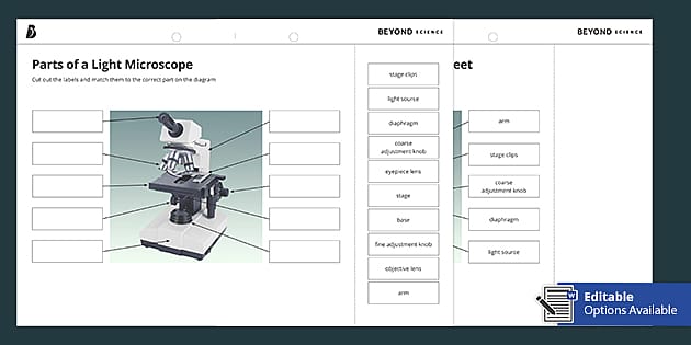
Parts of a Light Microscope Cut and Stick Worksheet - Twinkl
Compound Microscope Parts, Functions, and Labeled Diagram Compound Microscope Definitions for Labels. Eyepiece (ocular lens) with or without Pointer: The part that is looked through at the top of the compound microscope. Eyepieces typically have a magnification between 5x & 30x. Monocular or Binocular Head: Structural support that holds & connects the eyepieces to the objective lenses.

microscope drawing with label - Clip Art Library
Simple Microscope - Diagram (Parts labelled), Principle, Formula and Uses The working principle of a simple microscope is that when a lens is held close to the eye, a virtual, magnified and erect image of a specimen is formed at the least possible distance from which a human eye can discern objects clearly. Magnification formula The magnification power of a simple microscope is expressed as: M = 1 + D/F Where
Label the light microscope | Teaching Resources
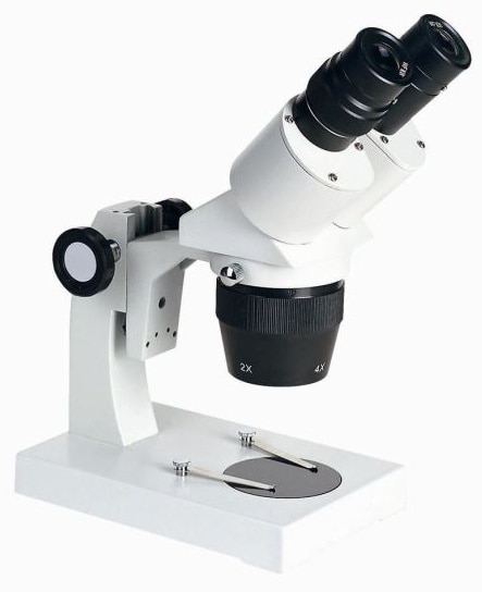
Parts of Stereo Microscope (Dissecting microscope) – labeled diagram ... Labeled part diagram of a stereo microscope ... (based on color bands and their respective labels), the objectives of a dissecting microscope are located in a cylindrical cone and, therefore, are not directly seen. ... but large enough to be seen or handled without the aid of a high power compound microscope. Thus, stereo microscopes have a ...
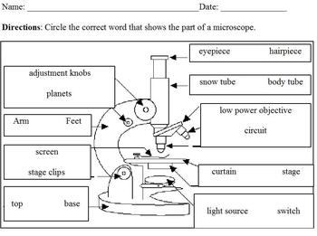
Microscope Labeling
Microscope Parts, Function, & Labeled Diagram - slidingmotion Microscope parts labeled diagram gives us all the information about its parts and their position in the microscope. Microscope Parts Labeled Diagram The principle of the Microscope gives you an exact reason to use it. It works on the 3 principles. Magnification Resolving Power Numerical Aperture. Parts of Microscope Head Base Arm Eyepiece Lens
Compound Microscope Parts, Functions, and Labeled Diagram ...
Wikipedia:Citation needed - Wikipedia To ensure that all Wikipedia content is verifiable, Wikipedia provides a means for anyone to question an uncited claim.If your work has been tagged, please provide a reliable source for the statement, and discuss if needed.. You can add a citation by selecting from the drop-down menu at the top of the editing box.In markup, you can add a citation manually using ref tags.

Compound Microscope Parts, Functions, and Labeled Diagram ...
How does a Microscope work A compound microscope has two or more lenses. The eyepiece or ocular lens sits atop the body tube. Many microscopes are binocular and have two ocular lenses. Additionally, a binocular head will have a prism, either in the head or the body tube, to split the image and direct it to both oculars.

Compound Microscope Parts – Labeled Diagram and their ...
Compound Microscope Parts, Functions, and Labeled Diagram Nov 18, 2020 · Common compound microscope parts include: Compound Microscope Definitions for Labels Eyepiece (ocular lens) with or without Pointer: The part that is looked through at the top of the compound microscope. Eyepieces typically have a magnification between 5x & 30x.

Label a Microscope Worksheet

Simple Microscope - Diagram (Parts labelled), Principle ...

The Microscope

How to use a Microscope - Microscopes 4 Schools
The Compound Light Microscope Label the following parts on ...

Microscope Components - Science Quiz

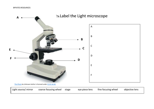
This is a common compound microscope. Label its parts from A ...

Microscope Labeling Activity - SMART Board Activity - Interactive Review
Microscope hi-res stock photography and images - Alamy
16 Parts of a Compound Microscope: Diagrams and Video ...

Compound Microscope Parts – Labeled Diagram and their ...
The Compound Light Microscope Label the following parts on ...
Introduction to the Microscope Lab Activity
Microscope Fill In The Blank - Fill Online, Printable ...
Simple Microscope - Parts, Functions, Diagram and Labelling ...

How to Use a Microscope (with Pictures) - wikiHow
National Middle School Standard Microscope, Monocular Head ...

Microscope Diagram - Free Printable Tests and Worksheets ...

Compound Light Microscope Labeling Diagram | Quizlet

Microscope Diagram Labeled, Unlabeled and Blank | Parts of a ...

Diagram of a Compound Microscope

Microscope Labeling

Microscope Diagram Labeled, Unlabeled and Blank | Parts of a ...
Compound Microscope Parts, Functions, and Labeled Diagram ...
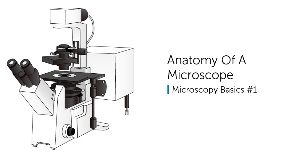
Anatomy Of A Microscope

Label the Microscope Diagram | Download Scientific Diagram

Biology label part of microscope

Lab Practical - Microscope parts and labelling Diagram | Quizlet
Simple Microscope - Diagram (Parts labelled), Principle ...
Post a Comment for "44 microscope diagram without labels"