42 heart structure and labels
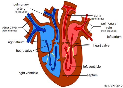
The Anatomy of the Heart, Its Structures, and Functions Heart Anatomy The heart is made up of four chambers: Atria: Upper two chambers of the heart. Ventricles: Lower two chambers of the heart. Heart Wall The heart wall consists of three layers: Epicardium: The outer layer of the wall of the heart. Myocardium: The muscular middle layer of the wall of the heart. Endocardium: The inner layer of the heart. Cells and cell structure quiz questions - Footprints-Science Biology random questions Cell structure Cell division Transport in cells Digestive system Heart and blood Health issues Plant tissues, organs and systems Communicable diseases Drugs Plant disease Photosynthesis Respiration Homeostasis Nervous system Hormones Reproduction Variation and Evolution Ecosystems Biodiversity Trophic levels Food production Chemistry …
Heart Anatomy Labeling Game - PurposeGames.com This is an online quiz called Heart Anatomy Labeling Game. There is a printable worksheet available for download here so you can take the quiz with pen and paper. Your Skills & Rank. Total Points. 0. Get started! Today's Rank--0. Today 's Points. One of us! Game Points. 19. You need to get 100% to score the 19 points available.
Heart structure and labels
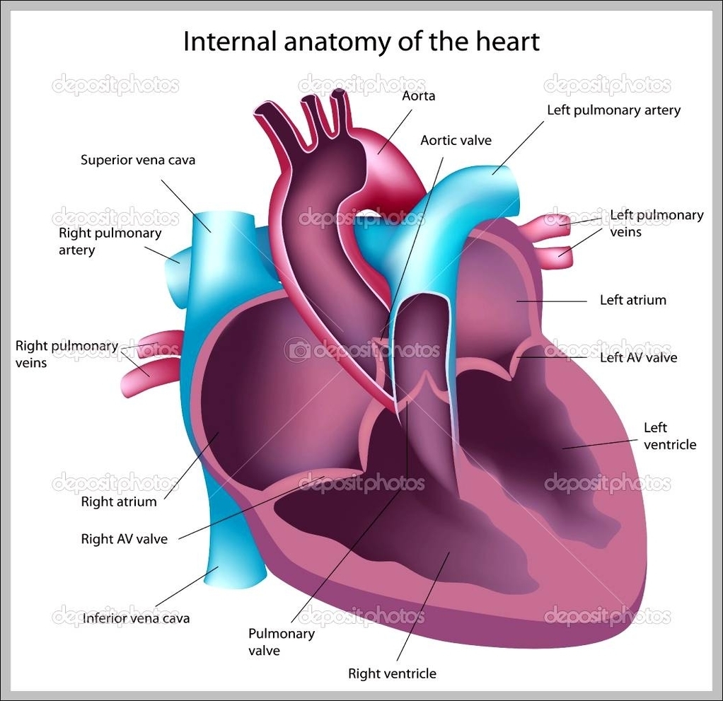
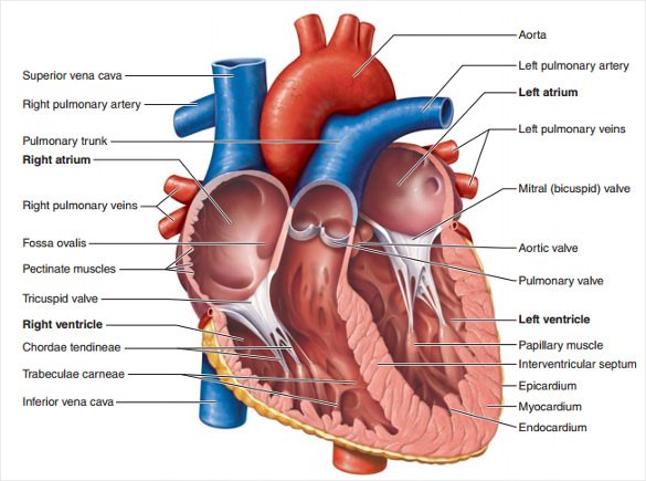
Dapagliflozin - Wikipedia Dapagliflozin, sold under the brand names Farxiga (US) and Forxiga (EU) among others, is a medication used to treat type 2 diabetes. It is also used to treat adults with certain kinds of heart failure and chronic kidney disease.. Common side effects include hypoglycaemia (low blood sugar), urinary tract infections, genital infections, and volume depletion (reduced amount of … Chapter 19: The Heart Flashcards | Quizlet •Allows heart to beat without friction, gives it room to expand and resists excessive expansion •Parietal pericardium-tough outer, fibrous layer of connective tissue-inner serous layer •Visceral pericardium (a.k.a. epicardium of heart wall)-serous lining of sac turns inward at base of heart to cover the heart surface Structure of the Heart | The Franklin Institute The two largest veins that carry blood into the heart are the superior vena cava and the inferior vena cava. They are called "vena cava" because they are the "heart's veins." The superior is located near the top of the heart. The inferior is located beneath the superior. A wall called a septum, separates the right and left sides of the heart.
Heart structure and labels. Human Heart Diagram Labeled | Science Trends List Of Heart Structures Heart Chambers Ventricles - The bottom two heart chambers. Atra - The upper two heart chambers. Wall Of The Heart Sinoatrial Node - A collection of tissue that releases electrical impulses and defines the rate of contraction for the heart. Atrioventricular Bundle - The fibers which transmit cardiac impulses. Heart Blood Flow | Simple Anatomy Diagram, Cardiac Circulation ... - EZmed Diagram: Blood flow through the right side of the heart involving the following cardiac structures: superior vena cava (SVC), inferior vena cava (IVC), right atrium (RA), tricuspid valve (TV), right ventricle (RV), pulmonary valve (PV), and main pulmonary artery (PA). Trick to Remember the Right Side Human Heart (Anatomy): Diagram, Function, Chambers, Location in Body The heart is a muscular organ about the size of a fist, located just behind and slightly left of the breastbone. The heart pumps blood through the network of arteries and veins called the... Structure of the Heart | SEER Training Structure of the Heart. The human heart is a four-chambered muscular organ, shaped and sized roughly like a man's closed fist with two-thirds of the mass to the left of midline. The heart is enclosed in a pericardial sac that is lined with the parietal layers of a serous membrane. The visceral layer of the serous membrane forms the epicardium.
The structure of the heart - Structure and function of the heart ... It is located in the middle of the chest and slightly towards the left. The heart is a large muscular pump and is divided into two halves - the right-hand side and the left-hand side. The... Heart Diagram with Labels and Detailed Explanation - BYJUS Well-Labelled Diagram of Heart The heart is made up of four chambers: The upper two chambers of the heart are called auricles. The lower two chambers of the heart are called ventricles. The heart wall is made up of three layers: The outer layer of the heart wall is called epicardium. The middle layer of the heart wall is called myocardium. Heart Labeling Quiz: How Much You Know About Heart Labeling? Here is a Heart labeling quiz for you. The human heart is a vital organ for every human. The more healthy your heart is, the longer the chances you have of surviving, so you better take care of it. Take the following quiz to know how much you know about your heart. Questions and Answers. 1. Heart Labels - Printable or Custom Printed Stickers | Avery.com Use our free specialty shape label templates to easily personalize your heart labels online. Customize one of our free designs or upload your own graphics and then choose the printing option that works best for you. Order your blank heart labels or custom printed heart labels and stickers online and get free shipping on orders of $50 more.
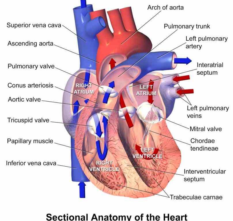
A Diagram of the Heart and Its Functioning Explained in Detail The heart blood flow diagram (flowchart) given below will help you to understand the pathway of blood through the heart.Initial five points denotes impure or deoxygenated blood and the last five points denotes pure or oxygenated blood. 1.Different Parts of the Body ↓ 2.Major Veins ↓ 3.Right Atrium ↓ 4.Right Ventricle ↓ 5.Pulmonary Artery ↓ 6.Lungs Ch. 19 Circulatory System- heart Flashcards | Quizlet Place the labels in order denoting the flow of blood through the pulmonary circuit beginning with the right atrium and ending in the left atrioventricular valve. The first and last structures are given. Right atrium 1. tricuspid valve 2. right ventricle 3. pulmonary valve 4. pulmonary trunk 5. pulmonary artery 6. lungs 7. pulmonary vein 8. left atrium Left atrioventricular valve. Place the ... PDF Free Anatomy Coloring Page - North Carolina State University The ate.2S the heart With oxygen ate labeled with at'l Color these areas The areas o' the heart with less oxygen ate labeled with a color areas BLUE. ARTERY LEFT LUNG PULMONARY VEINS AORTA PULMONARY VEINS raGHT LUNG ATRIUM RIGHT VENTRICLE INFERIOR VFNACAVA LEFT LEFT VENTRICLE AORTA BODY Downloaded from azcoloring.com 4 Song Structure Types to Know & When to Use Them in Your … 28.04.2022 · This is self-explanatory — the intro is the introduction to the song. And it’s one of the most important parts. According to Music Machinery, about 35% of listeners will skip a song within the first 30 seconds and nearly half of listeners skip a song before it’s over.That’s why your intro has to grab the listener’s ear and hold onto it.
Labelled Heart | Teaching Resources
Human Heart - Diagram and Anatomy of the Heart - Innerbody Because the heart points to the left, about 2/3 of the heart's mass is found on the left side of the body and the other 1/3 is on the right. Anatomy of the Heart Pericardium. The heart sits within a fluid-filled cavity called the pericardial cavity. The walls and lining of the pericardial cavity are a special membrane known as the pericardium.
37 Label The Anatomy Of The Heart - Labels 2021
How to Draw the Internal Structure of the Heart (with Pictures) To draw the internal structure of a human heart, follow the steps below. Part 1 Finding a Diagram 1 To find a good diagram, go to Google Images, and type in "The Internal Structure of the Human Heart". Find an image that displays the entire heart, and click on it to enlarge it. 2 Find a piece of paper and something to draw with.
13+ Heart Diagram Templates – Sample, Example, Format Download | Free & Premium Templates
Heart Anatomy: Labeled Diagram, Structures, Function, and Blood Flow There are 4 chambers, labeled 1-4 on the diagram below. To help simplify things, we can convert the heart into a square. We will then divide that square into 4 different boxes which will represent the 4 chambers of the heart. The boxes are numbered to correlate with the labeled chambers on the cartoon diagram.
called myocardium science External Structure Of Human Heart Anatomy structure of human heart ...
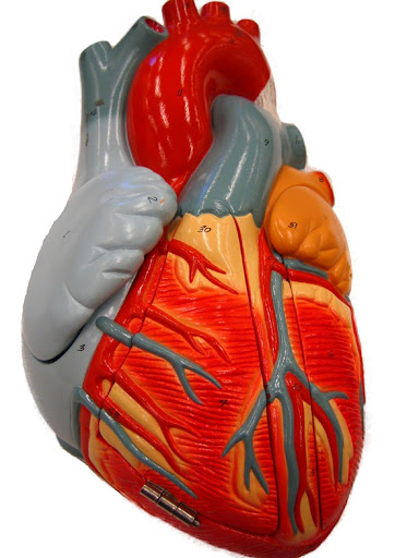
Human Heart Models | Heart Anatomy Models | Vitality Medical The heart model with labels is hand-painted with vivid colors to illustrate the papillary muscles, heart valves, and adjacent structures. show more Sort By Set Ascending Direction 4 Item (s) Show Magnetic Heart Model, Life Size, 5 Part G01 3B Scientific Starting at: $394.05 View Details Classic Heart Model 3B Scientific Starting at: $81.03
Know the structure of the heart - Labelled diagram
Flower Structure | BioNinja Flower Structures. The male part of the flower is called the stamen and is composed of:. Anther – pollen producing organ of the flower (pollen is the male gamete of a flowering plant); Filament – slender stalk supporting the anther (makes the anther accessible to pollinators); The female part of the flower is called the pistil (or carpel) and is composed of:
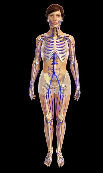
Female skeleton with backache — Stock Photo © kjpargeter #9310671
Heart: Anatomy and Function - Cleveland Clinic A layer of muscular tissue called the septum divides your heart walls into the left and right sides. Your heart walls have three layers: Endocardium: Inner layer. Myocardium: Muscular middle layer. Epicardium: Protective outer layer. The epicardium is one layer of your pericardium. The pericardium is a protective sac that covers your entire heart.

Label Function Human Heart Diagram And Function - Aflam-Neeeak
Labelling the heart — Science Learning Hub Blood transports oxygen and nutrients to the body. It is also involved in the removal of metabolic wastes. In this interactive, you can label parts of the human heart. Drag and drop the text labels onto the boxes next to the diagram. Selecting or hovering over a box will highlight each area in the diagram.
Free Unlabeled Heart Diagram, Download Free Clip Art, Free Clip Art on Clipart Library
Human Heart - Anatomy, Functions and Facts about Heart Label the Heart Diagram below: Practice your understanding of the heart structure. Drag and drop the correct labels to the boxes with the matching, highlighted structures. Instructions to use: Hover the mouse over one of the empty boxes. One part in the image gets highlighted.

Label The Heart Diagram - Human Anatomy
Structure Of The Heart | A-Level Biology Revision Notes The heart is divided into four chambers: The two atria (auricles): these are the upper two chambers. They have thin walls which receive blood from veins. The two ventricles: these are the lower two chambers. They have thick, muscular walls which pump blood through the arteries.
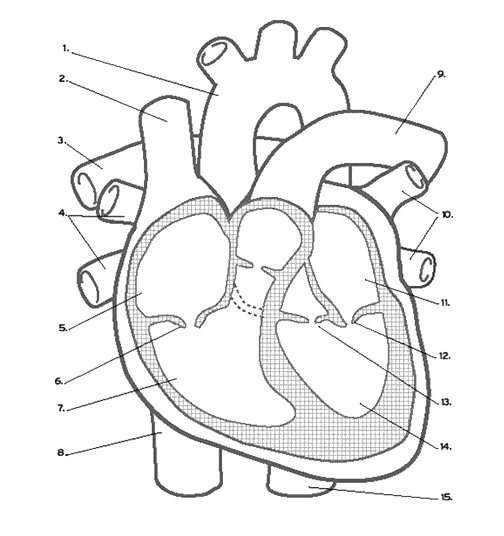
Heart Labeling (Internal)
The Heart - Science Quiz - GeoGuessr Aorta, Aortic valve, Left atrium, Left ventricle, Mitral valve, Pulmonary artery, Pulmonary valve, Pulmonary vein, Right atrium, Right ventricle, Septum, Superior vena cava, Tricuspid valve (13) Create custom quiz 0% | 0:05 | Click on: Aortic valve > Sound On Review

Circulatory System Diagram | New Health Advisor
Heart Diagram with Labels and Detailed Explanation The heart is located under the ribcage, between the lungs and above the diaphragm. It weighs about 10.5 ounces and is cone shaped in structure. It consists of the following parts: Heart Detailed Diagram Heart - Chambers There are four chambers of the heart . The upper two chambers are the auricles and the lower two are called ventricles.

38 Label The Following Diagram Of The Heart - Labels 2021
Label the heart — Science Learning Hub In this interactive, you can label parts of the human heart. Drag and drop the text labels onto the boxes next to the diagram. Selecting or hovering over a box will highlight each area in the diagram. Right ventricle Right atrium Left atrium Pulmonary artery Left ventricle Pulmonary vein Semilunar valve Vena cava Aorta Download Exercise Tweet

Eating for a healthy heart - Baker This may help to improve your overall heart health. Unhealthy ‘saturated and trans’ fats should be limited. This fact sheet will help you with: choosing healthy fats and sources; practical ways to include this into your diet; reducing unhealthy fats in your diet; how to read nutrition food labels. Download fact sheet
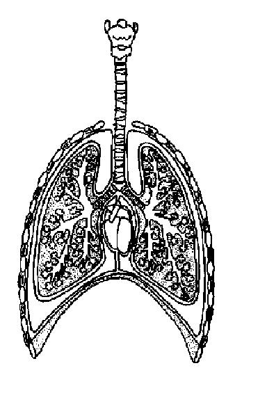
The Anatomy and Physiology of Animals/Respiratory System Worksheet - WikiEducator
Label Heart Anatomy Diagram Printout - EnchantedLearning.com Label Heart Interior Anatomy Diagram: Human Anatomy: The heart is a fist-sized, muscular organ that pumps blood through the body. Oxygen-poor blood enters the right atrium of the heart (via veins called the inferior vena cava and the superior vena cava). The blood is then pumped into the right ventricle and then through the pulmonary artery to ...
CLASS NOTES: Basic Heart Anatomy (Vital Signs: Understanding What the Body Is Telling Us)
Heart anatomy: Structure, valves, coronary vessels | Kenhub Heart anatomy. The heart has five surfaces: base (posterior), diaphragmatic (inferior), sternocostal (anterior), and left and right pulmonary surfaces. It also has several margins: right, left, superior, and inferior: The right margin is the small section of the right atrium that extends between the superior and inferior vena cava .

The Human Egg Cell Explained For Egg Donors - Altrui Egg Donation Agency
Heart Health | Heart Attack Prevention | Bayer® Aspirin Along with other heart-healthy choices, it can reduce your risk of having another heart attack. Learn About Aspirin's Benefits. Aspirin is not appropriate for everyone, so be sure to talk to your doctor before you begin an aspirin regimen. Surviving a heart attack can mean you’re at a higher risk for another. The good news: you can still manage other risk factors. GET PREVENTION …
Post a Comment for "42 heart structure and labels"